
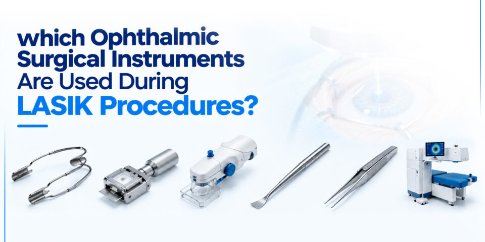
A LASIK procedure depends on five groups of ophthalmic surgical instruments: diagnostic devices, eyelid and eye stabilizers, flap-creation tools, the excimer laser, and hand instruments for flap handling. Diagnostic devices such as the pachymeter and corneal topographer determine whether an eye qualifies for surgery. The speculum and fixation forceps hold the eye steady during treatment. A femtosecond laser or a microkeratome creates the corneal flap, and the excimer laser then removes tissue to correct the refractive error. Flap lifters, forceps, and irrigation cannulas let the surgeon raise the flap and return it to position. Sterilization routines and calibration schedules decide how reliably the full set performs from one case to the next. At R L Hansraj Company, we supply instruments for each of these stages to surgeons and eye hospitals, and the sections below follow the order in which a surgeon uses them.
Diagnostic Devices That Decide LASIK Eligibility
Every LASIK case starts well before the operating room. Corneal topography maps the front surface of the cornea and identifies irregular astigmatism or early keratoconus, either of which rules out LASIK for that eye. A pachymeter measures corneal thickness. Surgeons generally want a residual stromal bed of at least 250 to 300 micrometres after the flap and the ablation, so the thickness reading sets the limit on how much tissue can be treated.
An aberrometer records higher-order aberrations and feeds wavefront-guided treatment profiles into the excimer laser. Autorefractors and a slit lamp complete the baseline work-up, and the slit lamp is used again after surgery to check flap position. Tear break-up time and Schirmer strips belong to this stage as well, because ocular surface disease affects both healing and the accuracy of corneal measurements.
These devices need scheduled calibration. A pachymeter that drifts out of range changes the safe ablation depth for a patient with a borderline cornea.
Eyelid Speculum and Fixation Instruments
The eyelid speculum keeps the lids apart for the full length of treatment. Wire speculums such as the Barraquer type give wide exposure with low pressure on the globe, while solid-blade designs hold the lashes away from the surgical field. Surgeons choose by lid anatomy. A narrow palpebral fissure calls for a different speculum than a deep-set eye, so most refractive suites keep several designs on the tray.
Fixation forceps grip the conjunctiva near the limbus to stabilize the globe when the surgeon needs added control. A corneal marker applies reference marks that guide flap realignment at the end of the case. The marker tip must be clean and dry before use, because smudged marks leave the surgeon unsure of the flap position.
Flap Creation: Femtosecond Laser and Microkeratome
Femtosecond Laser
A femtosecond laser delivers infrared pulses that form a plane of microscopic bubbles inside the cornea at a programmed depth. The surgeon sets the flap diameter, thickness, hinge position, and side-cut angle on the console. Flap thickness commonly falls between 90 and 120 micrometres. A suction ring and a disposable patient interface hold the eye against the applanating surface during the cut, which usually takes under a minute.
Microkeratome
A microkeratome uses an oscillating blade mounted on a head that travels across the cornea along a suction ring. Every case requires a new sterile blade, and the blade is inspected under magnification before use. A nick in the cutting edge produces an irregular flap surface and can lead to a poor visual result. The suction ring size and the depth plate control flap diameter and thickness.
Hand Instruments for Lifting and Repositioning the Flap
After the cut, the surgeon separates the flap edge from the stromal bed and folds the flap back along its hinge. Blunt-tipped designs lower the risk of tearing the flap margin. The same set of hand instruments handles the flap again once the laser treatment ends. Fine polished tips move across the stromal bed without scratching it, and rounded edges reduce pressure on the flap margin.
- Flap lifter or spatula: breaks the adhesion at the interface and raises the flap.
- Irrigating cannula: flushes debris and fluid from the interface after ablation.
- Cellulose sponges: dry the stromal bed before treatment, since stromal hydration changes the ablation rate.
- Flap-positioning spatula or sponge: aligns the flap to the reference marks and smooths out striae.
Once the flap is back in position, the surgeon waits a few minutes for adhesion and then checks for striae before removing the speculum. Wrinkles that go unnoticed at this point are harder to correct on a later visit.
The Excimer Laser Reshapes the Corneal Stroma
The excimer laser uses a 193-nanometre argon-fluoride beam to remove corneal tissue in layers a fraction of a micrometre thick per pulse. The wavelength breaks molecular bonds without heating the surrounding tissue. The treatment plan comes from the topography and aberrometry data collected during the work-up.
An eye tracker follows eye movement hundreds of times per second and repositions or pauses the beam when the eye shifts. Ablation time for a moderate prescription is often under a minute per eye.
Maintenance for this unit includes fluence checks and test ablations on calibration plates before each operating session. A laser that has not passed these checks should not be used on a patient.
Sterilization, Maintenance, and Instrument Sourcing
Reusable hand instruments go through cleaning, an ultrasonic bath, inspection under magnification, and steam sterilization according to the manufacturer’s instructions. Tips of lifters and cannulas show wear first. A burr on a spatula edge can damage the corneal epithelium, so any instrument with a bent tip, burr, or corrosion should be taken out of circulation.
Disposable items such as microkeratome blades and femtosecond patient interfaces are single-use and should never be reprocessed. Instrument logs that record sterilization cycle counts help your team decide when to replace reusable pieces.
When you equip a refractive suite, we at R L Hansraj Company help you match instrument specifications, tip profiles, and materials to your laser platform and your surgeons’ preferences.
Conclusion
A LASIK procedure relies on a defined sequence of equipment: diagnostic devices, eyelid and fixation instruments, a flap-creation system, an excimer laser, and a set of precise hand instruments. The reliability of these medical tools for surgery shows in repeatable flap thickness, accurate centration, and predictable healing. Calibration, inspection, and sterilization keep that performance consistent across cases.
If you are equipping or upgrading a refractive suite, contact R L Hansraj Company. We will review your platform and surgeon requirements and recommend the instruments that fit them.
Frequently Asked Questions
Which instruments create the corneal flap in LASIK?
A femtosecond laser or a microkeratome creates the flap. The femtosecond laser cuts with infrared pulses at a programmed depth, and the microkeratome cuts with an oscillating blade.
Is a femtosecond laser required for LASIK?
A femtosecond laser is not mandatory. Many surgeons cut the flap with a microkeratome, and the outcome depends on blade condition, suction stability, and the surgeon’s experience with the device. Femtosecond platforms give tighter control over flap thickness, which helps in thinner corneas.
Why is corneal thickness measured before LASIK?
The flap and the ablation both use up corneal tissue, and the remainder has to hold the eye’s shape for years. Surgeons plan so that roughly 250 to 300 micrometres of stromal bed stays behind. A cornea that measures thin at the first visit often points the patient toward PRK.
What does an eyelid speculum do during LASIK?
The speculum keeps the lids open for the whole treatment, so blinking cannot interrupt the laser. It also pulls the lashes away from the flap area, where they could carry debris onto the interface. A poorly fitted design that presses on the globe can make suction harder to maintain during the flap cut.
How often should reusable LASIK hand instruments be replaced?
No single interval applies to every instrument. Inspect each piece under magnification after every cleaning cycle, and replace any instrument with a burr, bent tip, or surface corrosion.
Which diagnostic instruments are needed before LASIK?
A corneal topographer, pachymeter, aberrometer, autorefractor, and slit lamp form the standard work-up. Tear film tests are added when the surgeon suspects dry eye.
Which LASIK instruments are single-use?
Microkeratome blades and femtosecond laser patient interfaces are single-use items. Sponges and similar disposables are also discarded after each eye.